Our Research
Developing protein-based tools to study neurotransmitter signaling in animals
Our brain has a language of its own. At the core of its complex functions are neurotransmitters, chemical messengers released between neurons to coordinate circuit activity. Understanding the chemical language of the brain is a goal of fundamental importance, as many of these signaling molecules or the cellular receptors that relay their signals, are involved in diseases of the nervous system and are potential targets of pharmaceuticals that could restore physiological brain functions. Towards this goal, an important first step is to decipher the associations between animal behavior, neural activity, and the precise spatial and temporal dynamics of these secreted molecules.
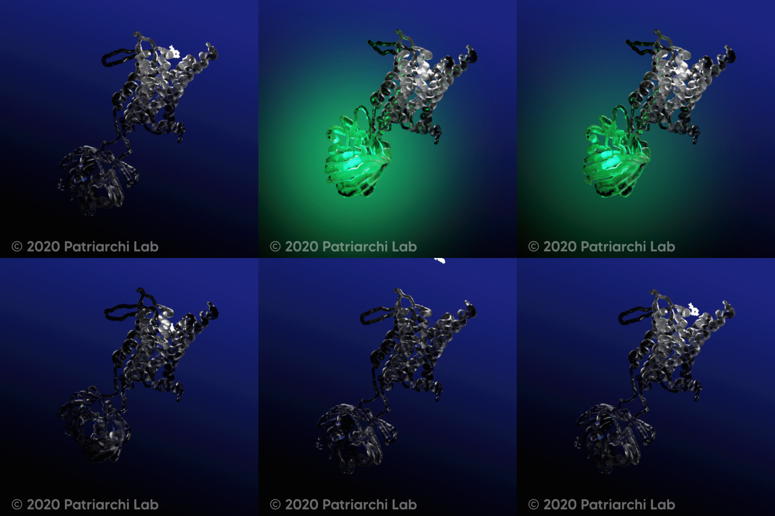
To aid our understanding of neural communication, along with continuous improvements in neuroimaging technologies, a range of new molecular tools needs to be developed and deployed. Genetically encoded fluorescent sensors, such as the widely utilized calcium sensors GCaMPs, occupy the center stage, due to their ideal properties for in vivo imaging and their flexible combination with the most advanced imaging modalities. Standing on the shoulders of these giants, recent developments by us and others led to the first genetically encoded indicators for dopamine (dLight1), a key neurotransmitter best known for its roles in reward and motivation. These highly sensitive sensors are built by engineering a single fluorescent protein (circularly permutated Green Fluorescent Protein) into G-protein coupled receptors (GPCRs).
We engineer new optogenetic tools and approaches
to investigate neurotransmitter signaling at the system level
Our team is interested in expanding and optimizing the neurotechnology toolbox, to shine a new light on the in vivo dynamics of diverse molecules involved in neural communication. Our ultimate goal is to leverage on the tools we build to unravel the neurotransmitter and receptor mechanisms of devastating psychiatric disorders such as depression. Because of the central pharmacological relevance of membrane receptors to human diseases, we also plan to deploy our new technologies for drug development.
Ongoing projects:
Our approach combines state of the art molecular biology techniques, custom-designed screening assays, ex vivo and in vivo brain imaging methods, automated behavioral tracking and mouse behavioral assays. We are also keen to establish cross-disciplinary collaborations involving the dissemination of our novel tools and expertise.
Our current projects focus on four main objectives:
1) Development of new genetically-encoded sensors and approaches for detecting neurotransmitters in vitro, ex vivo and in vivo with high sensitivity;
2) Development of new optogenetic tools and approaches for controlling endogenous receptor signaling;
3) Leverage on these tools for understanding the spatiotemporal coding of neuromodulatory signaling in the brain in healthy and diseased states;
4) Establish new platforms for ultra-high throughput screening of receptor-active molecules potentially useful for human disease treatment or prevention.
Allosteric modulation of a DA sensor
Here we developed a new approach for boosting the sensitivity of DA imaging with the biosensor dLight. This approach leverages on a highly specific molecule developed by Eli Lilly that acts as a positive allosteric modulator at the human dopamine D1 receptor. We thoroughly demonstrated that application of this molecule to the system under investigation (whether it be cultured DAergic neurons, brain slices, or living animals) enables flexible tuning of dLight sensitivity and allows us to track tonic versus phasic DA release properties on-demand.
NOPLight1
In this work we developed a new genetically-encoded sensor for detecting the opioid neuropeptide nociceptin (NOP, also known as orphanin-FQ peptide). We used the sensor to detect endogenous NOP release during acute stress and in assays to study the animal's motivation to consume a reward. This new tool will be useful to investigate how nociceptin release interplays with neural activity to constrain motivated and stress/anxiety behaviors.
nLights and other sensors
In this work we developed, characterized and validated new multicolor norepinephrine sensors, called nLightG and nLightR for the green and red sensor, respectively. This represents a new family of indicators based on an alpha-1 adrenergic receptor subtype. The sensors can detect norepinephrine in living animals with superior sensitivity, ligand specificity and temporal resolution as compared with previous tools. As part of this same work we developed new sensors for acetylcholine, histamine, adenosine, fractalkine.
GLPLight1 and Photo-GLP1
In this work we introduced a novel genetically encoded sensor based on the human GLP1 receptor, which we named GLPLight1. We demonstrate that its fluorescence signal accurately reports the expected efficacies and potencies of different pharmacological ligands, with very high sensitivity and temporal resolution. Using this sensor, we established an all-optical assay to characterize a novel photocaged GLP-1 derivative (photo-GLP1) and to demonstrate optical control of GLP1R activation. Thus, the new all-optical toolkit introduced here enhances our ability to study GLP1R activation with high spatiotemporal resolution. Read more here: https://elifesciences.org/articles/86628#annotations
Make it stand out
Photo-OXB
We introduced the first photocaged orexin-B derivative (photo-OXB). Since OXB lacks functionalizable amino acids in its biologically relevant C-terminal region, we developed an approach for C-terminal photocaging of peptides, which should be directly applicable to other peptides that activate their cognate receptor via their C terminus. We demonstrated the compatibility between optical uncaging of OXB and biosensor imaging and used all-optical assays to functionally characterize photo-OXB light sensitivity and bioactivity. Finally, we showed that photo-OXB can be used for light-controlled activation of endogenous orexin receptors ex vivo by demonstrating the effect of its uncaging on the membrane potential of medium spiny neurons in acute brain slices. We expect that the photo-OXB tool described here will be rapidly adopted to investigate the multifaceted functions of orexin signaling in physiological or disease contexts. In addition, the caging strategy we developed, in combination with optical biosensor assays, will be a valuable resource for the broader chemical biology community as it allows for rapid development of photocaged peptides and their optical characterization. Read the article here: https://www.cell.com/cell-chemical-biology/fulltext/S2451-9456(22)00413-5
OxLight1
In our recent work, we leveraged on our experience in protein engineering to develop a new genetically encoded biosensor, named OxLight1, that is capable of detecting orexin neuropeptides (also known as hypocretins). This sensor is based on a circularly permuted green fluorescent protein integrated into the human type-2 orexin receptor. Compared to the previous screening-intensive efforts deployed for the development of dLight or the GRAB family of neuromodulator sensors, in this work we demonstrated a more efficient tailored screening approach for sensor engineering focused on the insertion/deletion or mutagenesis of individual GPCR residues at a time. With this method we could efficiently optimize a GPCR-based sensor with exceptional sensitivity (maximal fluorescence response of >8-fold) with just 100 variants. We demonstrated that OxLight1 responds to both orexin-A and B with nanomolar affinity, sub-second activation kinetics and high molecular specificity without noticeable coupling to endogenous cellular, making this an ideal tool for probing endogenous orexin release in living animals. Read the article here: https://www.nature.com/articles/s41592-021-01390-2


